Past is Prologue
The skeletal collections that Carlina de la Cova has relied on for forensic techniques have their own stories to tell.
A lot of people have contributed to the creation of modern forensic science — and many of them did so unwittingly. Anthropologist Carlina de la Cova is part of a new generation of forensic scientists who want to do something about that.
De la Cova’s research in biological anthropology often involves working with skeletal collections that were typically put together during the first half of the 20th century. She has made extensive use of the Hamann-Todd collection, for example, which comprises more than 3,100 individual skeletons and was assembled by professors of anatomy at what is now Case Western Reserve University. The skeletons were prepared from cadavers starting in 1893 and are now held in the Cleveland Museum of Natural History.
They’re not skeletons, they’re people to me because for some of them I know their stories. I feel it’s my duty to tell the story of these individuals.
—Carlina de la Cova
According to de la Cova, Hamann-Todd and similar collections were crucial to the development of modern forensic science.
“These skeletons have given us the basic human identification techniques in our discipline,” de la Cova says. “They have been used to create the methods to identify sex, to identify age, to assess ancestry in this country from skeletal remains.”
De la Cova knows about those techniques firsthand, using them in the field in her position as a deputy coroner for Richland County and as a forensic consultant in other states.
The osteological collections that have proven to be indispensable to her discipline and to the detectives she supports do not, however, contain the remains of civic-minded individuals who donated their bodies to science. Rather, they represent in large part the final resting place of people of little means who died far from home.
In the collections de la Cova has studied, bodies that were not claimed by relatives within 24 to 32 hours were sent to medical schools for dissection, she says. Some of those were then selected by the anatomists for preservation in their collections.
De la Cova has spent time further identifying the individuals, working from their names and the hospitals from which they originated.
“They were indigent, they were immigrants, they were migrants,” she says. “In fact, a lot of the African-Americans I’m finding in these collections were part of the Great Migration.”
The first wave of that large-scale movement of black Americans out of the South took place roughly between 1910 and 1930. “I’m finding that 32 to 40 percent of the white females, in the collections I’ve worked with, were in mental institutions,” she says.
De la Cova is burnishing the dataset of these canonical collections, adding information about individuals, such as institutionalization status or medical histories, that perhaps ought be taken into account when forensic scientists estimate things like height, weight or age from skeletal observations or measurements. She thinks there might be fine details in individual skeletal histories that have the potential to make forensic predictions even more precise.
But she’s also trying to develop with her colleagues a better sense of how to ethically work with the collections.
“They’re not skeletons, they’re people to me because for some of them I know their stories,” de la Cova says. “I feel it’s my duty to tell the story of these individuals.
“In the early history of the discipline, they were objects to be studied, to be objectified. The newer generation of bioarchaeologists, of biological anthropologists, are pushing beyond that,” she says. “The folks that we study, we have to think about who their descendants are, and we have to actively engage in discussions with their descendants. That’s the way the discipline is now moving.”
Black Death
Why a horrific disease outbreak from more than 600 years ago might help us prepare for future outbreaks.
An investigator approaches the workbench, opens a box, removes a pelvis, a skull, arm and leg bones, and other parts of a mostly complete skeleton. She makes a series of measurements and observations: femur length, hip shape, tooth development and many more.
She examines each bone for clues. Was there an earlier fracture? Evidence of disease? Any signs of stress early in life?
She’s collecting these data points to build as complete a profile as possible of the person whose skeleton she is examining: age, sex, stature, health status, whether the cause of death can be specified, and so on.
So what kind of investigator is this? Forensic technician? Forensic pathologist? Forensic anthropologist?
In Sharon DeWitte’s case, it’s none of the above. She is an anthropologist, specializing in biological anthropology, but you can’t find the word forensic anywhere on her vita.
I definitely think that, even in the case of something like Ebola, we can learn from past epidemics. There are mechanisms that we might put into place to prevent the kinds of levels of mortality that we’re seeing.
—Sharon DeWitte
The adjective “forensic” indicates that the noun it modifies has something to do with the court system. That could mean a criminal case, or it could mean a legal determination about property rights involving remains in a cemetery. In DeWitte’s work, it doesn’t apply. She wants to use the tools of modern anthropology to understand the lives of people who died a long time ago rather than take on forensic cases.
“I made a decision when I was in graduate school to work with remains that are the least contentious possible, so I actually do not work with Native American remains, for that exact reason,” DeWitte says. “I don’t think there’s anything that I can do that would trump the wishes of descendant populations, so instead, I work with dead British people.”
Understanding those dead British people requires all the skills of a forensic investigator — skills that she helps teach the next generation of forensic scientists in the classroom. And she uses them as a matter of routine in studying people who died hundreds of years ago.
DeWitte’s research focuses on the Black Death, the pandemic that ravaged London and most of the inhabited world in the 14th century, killing tens of millions and possibly more than half of Europe’s population in just a few years.
That was a long time past, in a place far removed from the modern world, but DeWitte is drawing conclusions about pandemics that are relevant today, she says. The Black Death, for example, with a more than 50 percent fatality rate, was long thought to be an indiscriminate killer. If you came down with it, it was just a roll of the dice whether you survived. But DeWitte’s research has shown that the dice were, in fact, loaded: the Black Death was more virulent with children, the elderly and the infirm.
It’s an insight that policymakers would be advised to bring to bear on modern-day outbreaks, DeWitte says.
“By demonstrating that people varied in their risk of death during the Black Death, in what was one of the worst pandemics in human history, we should expect those same sort of patterns regardless of the cause of mortality,” she says. “I definitely think that, even in the case of something like Ebola, we can learn from past epidemics. There are mechanisms that we might put into place to prevent the kinds of levels of mortality that we’re seeing.
“I actually mentioned this in a recent paper. The recent outbreak of Ebola killed 10 times as many people as all of the previous outbreaks combined, going back to 1975. The genetic variant that caused this outbreak was the same as previous outbreaks, the case fatality ratios were the same, the transmissibility was the same and the symptoms were the same. It also had a much wider geographical distribution than any previous outbreak, and that is entirely the result of human behavior. It was just that people engaged in behaviors that completely promoted the spread of the disease.”
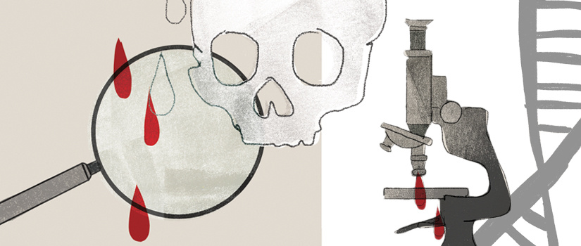
Steaming out Luminol’s Wrinkles
A new technique called ‘steam thermography’ could replace a traditional chemical
in detecting blood at crime scenes.
Luminol, a chemical used to detect blood, gets trotted out on TV crime shows, but a new technique might someday compete with the storied forensics tool as a means of solving real crimes.
Carolina chemistry professors Michael Myrick and Stephen Morgan are showing that what they call “steam thermography” has the ability to spot blood spots where luminol can’t.
Myrick and Morgan use a hand steamer in combination with thermal imaging in their new visualization method, which can make even a highly diluted blood sample stand out from its background. Their laboratory work has roots in the technology of night-vision goggles, which contain an infrared sensor that differentiates between areas from which large and small amounts of IR radiation, commonly referred to as heat radiation, are emanating. The device converts the data into a visible image distinguishing between warm and cool areas — perfect for making a hot-blooded creature stand out from the background.
Myrick, Morgan and their students have been carrying that idea further, working to develop IR-sensitive cameras into instruments that highlight more than the difference between warm and cold. The light radiation that any object emits is a function of the molecules that make it up, so the research team has been refining the ability of IR-sensitive cameras to recognize chemical contrast; to “see” the difference between chemically distinct objects that are otherwise indistinguishable in an image.
Exhibit A in that effort is blood. Using grant money from the National Institute of Justice, the researchers are developing a system to make blood spots stand out from the background in a thermal image.
In the course of trying different approaches to enhance contrast, they found one that was eye-popping. By first treating a piece of blood-spattered fabric with a hand steamer, the team showed that droplets of even 1/1,000-diluted blood would stand out brightly in thermal imaging under the right conditions. And the contrast enhancement is evident in real time — an observer can watch blood spots emerge on the IR camera’s viewscreen as steam is applied.
The team has worked with trained forensic scientists who verified that highly diluted bloodstains that their “steam thermography” technique reveals are not visible with standard light visualization.
That would make this one of the few techniques to give luminol a run for its money. Luminol is not used as extensively in crime labs as its portrayal in on TV might imply. Luminol and a reagent must be sprayed on a surface of interest, which effectively contaminates a crime scene.
As a consequence, using luminol is often a last resort, when other methods fail to highlight where blood is present — when someone tried to clean it up, for example, and not much is left. With demonstrated success at 1/1000 dilution, steam thermography is a promising alternative.
What’s more, you need a darkened room to see the faint, distinctive blue glow that is generated for a short time when the luminol solution has come in contact with blood. That precludes its use outside. Steam thermography doesn’t have that limitation.
Myrick and Morgan’s method does introduce a brief steam exposure to a crime scene, but they show that adding an already ubiquitous chemical (water) that quickly evaporates doesn’t warm the surface much (less than 20 degrees Fahrenheit), and they have verified that DNA testing of blood spots wasn’t affected.
Moreover, they’ve also established that their new technique isn’t fooled by common materials that can cause false positives with luminol, which include bleach, rust and coffee stains. That makes steam thermography a good candidate for filling some niches where luminol doesn’t fit — and perhaps even some where it does.
3-D Printing a Crime Scene
An engineering student’s CAD skills might help save a death row inmate’s life.
Success in the engineering laboratory opened an unexpected door to the world of forensic science for one undergraduate at Carolina. Shana Mussel, who will earn her bachelor’s degree later this year, excelled in computer assisted design (CAD) in her mechanical engineering curriculum. It was a skill that her professor Joshua Tarbutton knew would carry her to success in a forensic reconstruction project he agreed to oversee last summer.
Through connections in the community, Tarbutton had learned about a death row inmate, near the end of his appeals, who needed help in the courtroom. Convicted of murdering a police officer more than 20 years ago, the inmate has been trying to establish his contention that he did not know he was being confronted by a police officer and had been startled at the time of the shooting. He tried to reconstruct the shooting scene with a model to make the point, but the cardboard contraption he had assembled didn’t do much to strengthen his argument.
Possibly saving a man’s life, through being an engineer — that’s pretty cool. Definitely not something you think you’ll be doing with this kind of degree.
—Shana Mussel
At the behest of Justice 360, a non-profit that provides quality legal representation to death row inmates, Tarbutton enlisted Mussel to re-create the scene of the crime. Using forensic photographs and other evidence, she modeled the house and porch from which the police officer was shot using Sketchup, a three-dimensional modeling platform to which her CAD skills were readily transferred. They then brought the scene to life through 3-D printing, generating a scale model of the house, its porch and the two men involved.
Tarbutton and Mussel then prepared a report based on the geometry of the shooting established by the evidence. It showed that the inmate’s view of the officer was obscured when he fired, and that his gun was fired from the hip, not the shoulder.
Those conclusions could make a difference for a man whose life literally hangs in the balance, and a hearing on the case is expected later this year. If a new trial takes place, both engineers likely will testify.
Mussel won’t appear as a surprise witness in court by any means, except perhaps to herself. “Possibly saving a man’s life, through being an engineer — that’s pretty cool,” Mussel says. “Definitely not something you think you’ll be doing with this kind of degree.”
You Can’t Hide Your Lyin’ Mind
A psychologist uses fMRI imaging to detect the moment of deception.
The lie detector test, or polygraph, is viewed skeptically in the courtroom, suspect enough to be subject to a patchwork of federal, state and local laws about whether the test results can even be admitted as evidence.
Psychology professor Jennifer Vendemia and graduate student Jimmy Nye hope that the new scientific tools they’re bringing to bear in the study of deception will make lie detection a more reliable courtroom witness in the next decade or so. They and their colleagues are using the tools of neuro-science to get an inside look into the mind as it seeks to deceive — something that takes a lot of brainpower not just in the thinking, but also in the doing, and with possible consequences for telling the truth.
To get a clear view of the lying mind, the researchers rely on two tools that give somewhat complementary information, the electroencephalogram (EEG) and functional magnetic resonance imaging (fMRI). An EEG measures how electrical impulses travel along the scalp, reflecting electrical flow between parts of the brain near the skull, and it can resolve brain activity on a microsecond time scale.
This is still preliminary, but what we’re starting to see is that the more lies you tell doesn’t really change how hard a lie was. What really changes, though, the more lies you tell, is that it makes it harder to tell the truth.
—Jimmy Nye
The fMRI technique provides much more spatial information, pinpointing in three-dimensional detail the blood flow within the brain, but the time scale cannot be resolved nearly as precisely as with the EEG. An fMRI image is only available every several seconds and reflects a time-averaged picture of blood flow over those several seconds.
Together, these tools open a new window onto the act of deception. Today’s polygraph measures only the after-effects of telling a lie — physiological responses such as changes in heart rate, breathing and sweating. With EEG and fMRI, Vendemia and Nye are able to watch the formulation of the intent to lie, which they have shown can often take a lot more work than telling the truth.
A truth-teller largely accesses parts of the brain associated with memory, presumably to recall the facts requested. Someone intent on deception, however, has a more complex response, accessing other parts of the brain that might be involved in processing the lie to ensure consistency or in calming the emotional responses that most people have to intentionally being untrustworthy.
Even the act of speaking an untruth is difficult. “We’ve found that the motor part of the lie, the actual telling of it, can take more time than truth-telling,” says graduate student Jimmy Nye. “We’ve seen that from measuring reaction times after a question was asked.”
What’s more, a person telling a series of lies appears to have increasing difficulty in unexpected ways.
“This is still preliminary, but what we’re starting to see is that the more lies you tell doesn’t really change how hard a lie was,” Nye says. “What really changes, though, the more lies you tell, is that it makes it harder to tell the truth.”
Having to maintain a lie throughout an interrogation might thus be reflected in difficulty in honestly answering even innocuous questions, which could be quantitatively detectable by a forensic examiner. “That’s something we really want to test, these long-term lies,” Nye says. “That’s where I see this going.”